How to Read Your Dental X‑Ray and 3D Scan: A Simple Guide for Gurgaon Patients
A clear, patient-friendly guide to common dental X‑rays and CBCT/3D scans used for implants, root canals and orthodontics, with practical tips on what images show and the questions to ask your dentist in Gurgaon.

Why dental images matter
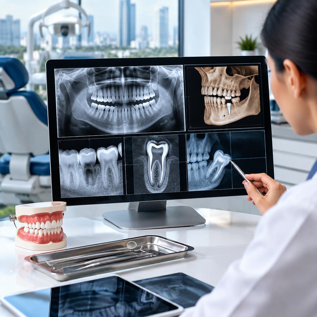
Dental X‑rays and 3D (CBCT) scans give your dentist views you can’t see with the naked eye. They help detect cavities between teeth, assess bone support, locate infections at a tooth’s root, and plan treatments like implants, root canals, and orthodontics. Clinics in Gurgaon, including modern multi‑specialty centres, commonly combine digital intraoral X‑rays with CBCT when complex planning or precise surgical guidance is needed.
The common X‑rays you’ll see
There are a few routine 2D X‑rays: bitewings (small, focused images of upper and lower teeth used to spot interproximal cavities and evaluate restorations), periapical films (show an entire tooth from crown to root and the surrounding bone—useful for root canal or infection assessment), and panoramic X‑rays (a broad overview of the jaws that shows wisdom teeth, the jaw joint, and general bone contours). Each has a different purpose and is chosen based on the problem being investigated.
What CBCT (3D) scans show
A CBCT or 3D scan produces cross‑sectional images of bone, teeth, and anatomical structures. For implant planning it reveals bone height, width, and the position of nerves or the sinus. For endodontic (root canal) cases it can show complex root anatomy and hidden fractures. In orthodontics it helps evaluate tooth positions and jaw relationships. CBCT gives a spatial map your dentist can use for safer, more predictable treatment planning.
How common problems appear on images
Some visual cues are useful to know—cavities often appear as darker areas in tooth enamel or between teeth on X‑rays; dense structures such as fillings, crowns and bone appear lighter. Bone loss from gum disease shows as reduced height of the bone around tooth roots and less dense bone patterns. An infected root tip typically shows as a small dark area near the root end. Impacted wisdom teeth are visible on panoramic images as teeth trapped under bone or pressing on adjacent roots. These are signs your dentist will interpret alongside clinical examination—avoid self‑diagnosis.
Radiation and safety (what patients should ask)
Digital X‑rays use very low radiation; CBCT involves higher dose than a single intraoral film but is still controlled and used only when the diagnostic benefit outweighs the exposure. Ask your dentist why a particular scan is needed, whether a lower‑dose option is available, and how the image will change your treatment plan. Clinics that emphasise modern imaging typically follow safety protocols and explain risks clearly—patients often report feeling reassured when the team discusses these details before scanning.
Questions to bring to your appointment
To get the most from your imaging review, ask: Can I see the images and have them explained? What do the darker or lighter areas mean for my teeth and bone? Is a CBCT necessary for my case, and how will it change treatment? Are there alternatives to the proposed procedure? What are the next steps and expected timeline? If you’re anxious about a procedure, ask about comfort options—many clinics in Gurgaon offer sedation and clear post‑op instructions.
How images guide implants, RCTs and orthodontics
For implants, images confirm if there’s enough bone and where to place the implant relative to nerves and sinuses; 3D planning reduces surprises during surgery. For root canals, periapical films and 3D scans reveal root shape and hidden canals to help the specialist clean and seal them. For orthodontics, digital scans and panoramic images show developing teeth, spacing and impacted teeth that affect the treatment plan. Having the right image at the right time improves predictability and long‑term results.
After the scan: records and second opinions
Ask for a copy of your digital images and a concise report—these are useful for tracking progress or seeking a second opinion. Keep prior X‑rays when starting a multi‑step treatment so your dentist can compare changes over time. Many patients appreciate clinics that combine advanced imaging with clear explanations and a comfortable environment; if you’re unsure, bring your images to a follow‑up consult to review options calmly and make an informed decision.
Customer review


Article by
Cosmodontist Dental Clinic 65
Cosmodontist Dental and Implant Centre, located in Sector 65, Gurgaon, is regarded as one of the Best Dental Clinics in Gurgaon. Headed by Dr. Nlay AB and Dr. Vidushii Mayor, the clinic is trusted by patients for expert care and advanced treatments. We aim to make world-class dentistry accessible by offering high-quality treatments at affordable prices. Whether you are searching for a dentist near me for routine check ups or full mouth rehab, CDC ensures lasting results. Our services include dental implants, painless root canal treatment, smile makeovers, Invisalign, instant teeth whitening, and cosmetic dentistry. With award-winning recognition and patient trust, Cosmodontist stands as a top dental clinic in Gurgaon.